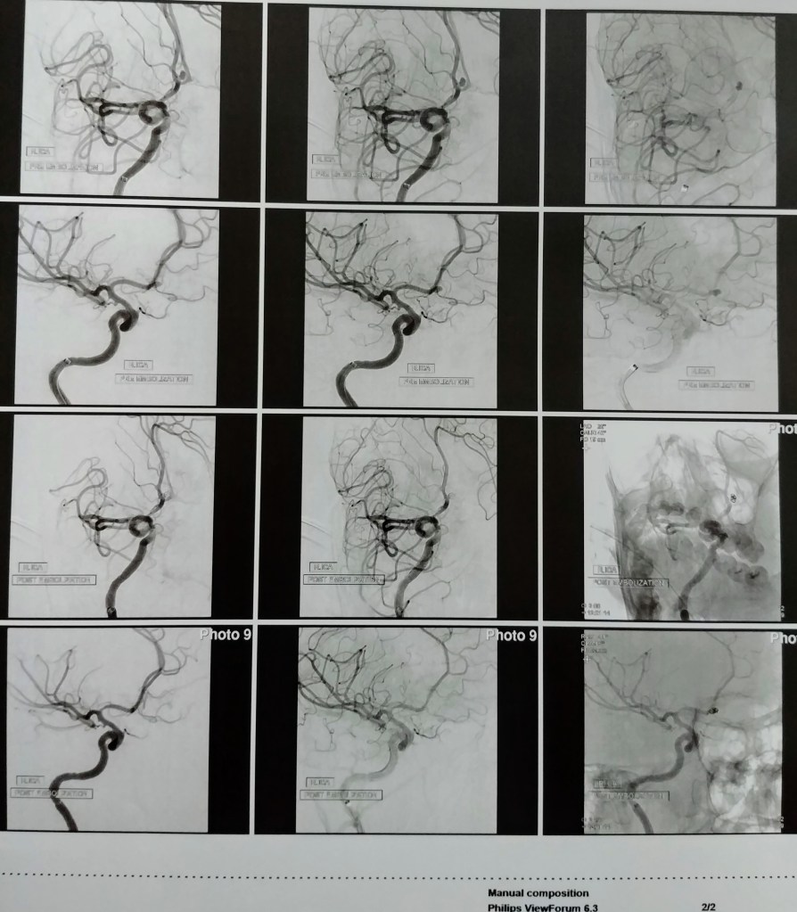
Can you spot the 2 aneurysms? The big balloon was the largest and below it was the smallest one that is still intact. This is one of the pictures from my angio showing the aneurysm before the embolization. Zoom in close for details.This scan compares the brain pre-embolization (top half) and post-embolization (bottom half). Zoom in close for a clear view and details.
If you have experienced a severe headache or have any other symptoms related to a ruptured aneurysm your doctor will order tests to determine if blood has leaked into the space between the skull bone and brain.
Several tests are available to diagnose brain aneurysms and determine the best treatment. These include:
Computed tomography (CT). This fast and painless scan is often the first test a physician will order to determine if blood has leaked into the brain. CT uses x-rays to create two-dimensional images, or “slices,” of the brain and skull. Occasionally a contrast dye is injected into the bloodstream prior to scanning to assess the arteries, and look for a possible aneurysm. This process, called CT angiography (CTA), produces sharper, more detailed images of blood flow in the brain arteries. CTA can show the size, location, and shape of an unruptured or a ruptured aneurysm.
Magnetic resonance imaging (MRI). An MRI uses computer-generated radio waves and a magnetic field to create two- and three-dimensional detailed images of the brain and can determine if there has been bleeding into the brain. Magnetic resonance angiography (MRA) produces detailed images of the brain arteries and can show the size, location, and shape of an aneurysm.
Cerebral angiography. This imaging technique can find blockages in arteries in the brain or neck. It also can identify weak spots in an artery, like an aneurysm. The test is used to determine the cause of the bleeding in the brain and the exact location, size, and shape of an aneurysm. Your doctor will pass a catheter (long, flexible tube) typically from the groin arteries to inject a small amount of contrast dye into your neck and brain arteries. The contrast dye helps the X-ray create a detailed picture of the appearance of an aneurysm and a clear picture of any blockage in the arteries.
Cerebrospinal fluid (CSF) analysis. This test measures the chemicals in the fluid that cushions and protects the brain and spinal cord (cerebrospinal fluid). Most often a doctor will collect the CSF by performing a spinal tap (lumbar puncture), in which a thin needle is inserted into the lower back (lumbar spine) and a small amount of fluid is removed and tested. The results will help detect any bleeding around the brain. If bleeding is detected, additional tests would be needed to identify the exact cause of the bleeding.
Brain aneurysms/cerebral aneurysms form when the walls of the arteries in the brain become thin and weaken. Aneurysms typically form at branch points in arteries because these sections are the weakest. Occasionally, cerebral aneurysms may be present from birth, usually resulting from an abnormality in an artery wall. Reference
Risk
factors for developing an aneurysm
Sometimes
cerebral aneurysms are the result of inherited risk factors, including:
genetic
connective tissue disorders that weaken artery walls
polycystic
kidney disease (in which numerous cysts form in the kidneys)
arteriovenous malformations (snarled
tangles of arteries and veins in the brain that disrupt blood flow. Some
AVMs develop sporadically, or on their own.)
history
of aneurysm in a first-degree family member (child, sibling, or parent).
Other
risk factors develop over time and include:
untreated
high blood pressure
cigarette
smoking
drug
abuse, especially cocaine or amphetamines, which raise blood pressure to
dangerous levels. Intravenous drug abuse is a cause of infectious mycotic
aneurysms.
age
over 40.
Less
common risk factors include:
head
trauma
brain
tumor
infection
in the arterial wall (mycotic aneurysm).
Additionally,
high blood pressure, cigarette smoking, diabetes, and high cholesterol puts one
at risk of atherosclerosis (a blood vessel disease in which fats build up on
the inside of artery walls), which can increase the risk of developing a
fusiform aneurysm.
Risk
factors for an aneurysm to rupture
Not all
aneurysms will rupture. Aneurysm characteristics such as size, location,
and growth during follow-up evaluation may affect the risk that an aneurysm
will rupture. In addition, medical conditions may influence aneurysm rupture.
Risk
factors include:
Smoking.
Smoking is linked to both the development and rupture of cerebral aneurysms.
Smoking may even cause multiple aneurysms to form in the brain.
High
blood pressure. High blood pressure damages and weakens arteries, making
them more likely to form and to rupture.
Size.
The largest aneurysms are the ones most likely to rupture in a person who
previously did not show symptoms.
Location.
Aneurysms located on the posterior communicating arteries (a pair of arteries
in the back part of the brain) and possibly those on the anterior communicating
artery (a single artery in the front of the brain) have a higher risk of
rupturing than those at other locations in the brain.
Growth.
Aneurysms that grow, even if they are small, are at increased risk of rupture.
Family
history. A family history of aneurysm rupture suggests a higher
risk of rupture for aneurysms detected in family members.
The
greatest risk occurs in individuals with multiple aneurysms who have already
suffered a previous rupture or sentinel bleed.
For my case, I had been diagnosed
with high blood pressure in the first trimester of my second pregnancy and the
condition was being managed when I suffered the brain aneurysm. No family
history of brain aneurysms that I am aware of. No smoking, no alcohol abuse,
and absolutely no drug use except for the ones for BP and vitamins. My take
from this is that we are all pretty much at risk. I have heard stories of
completely healthy people, very athletic, and with no family history get them. The
more information we know about brain aneurysms, the more lives we can help
save.
Let’s be honest, how many people are familiar with brain
aneurysm? Even with a background in healthcare, I did not know what brain aneurysms
were all about except for the definition. Often, we find ourselves becoming
quite familiar with a condition once we experience it either directly or
indirectly.
Think of a weak spot in a balloon and how it feels stretched
out and thin. A brain
aneurysm is like that. It’s a weak spot in the wall of a blood vessel
inside the brain.
That area of the blood
vessel gets worn out from constant flow of blood and bulges out, almost like a
bubble. It can grow to the size of a small berry.
Although brain
aneurysms sound alarming, most don’t cause symptoms or health problems. You can
enjoy a long life without ever realizing that you have a brain aneurysm.
But in rare cases, aneurysms can grow big, leak, or explode.
Bleeding
in the brain, known as hemorrhagic stroke, is very serious and
requires urgent medical care.
A ruptured brain aneurysm can be life-threatening and lead
to:
cerebral vasospasm (reduced blood flow to the brain)
I remember having a constant headache for 2 days or so prior to the rupture. The pain wasn’t overwhelming and I could tolerate it. I would rate it a 2 on the scale of 1-10 with 10 being the worst. New mothers, me being one of them, tend to ignore little things that their bodies are trying to communicate. We give excuses in order to make ourselves feel better. It is very true we are tired most of the time with little to no adequate rest most of the time. We prioritize the needs of our children and our loved ones. The question that still ponders my mind is – ‘was the aneurysm ruptured already prior to my hospital admission and the bleeding just got worse or did it rupture on that hectic night?’ The doctors that I have met with haven’t given me a conclusive answer yet. They tend to say…..in so many words——“we just don’t know”
So what are the actual symptoms of a brain aneurysm?
Please do not sit and start guessing what could be wrong with you or your loved one. Get emergency care if you suddenly get an intensely painful headache, lose consciousness, or have some of these other symptoms of an aneurysm that has ruptured: I cannot emphasize enough to you how critical it is to get that medical care urgently. Get help if you think something is wrong with your body. DO NOT WAIT.
Although brain aneurysms usually don’t show symptoms, they can press on the brain and nerves as they get bigger. See a doctor at once if you’re having the following symptoms of an unruptured aneurysm:
Please remember that no matter what you are going through or how things turn out, you are not alone. God is with you and He has everything under control. He will lead you to where you need be at the right time. He will bring the right people to your case. God is watching us from a distance. You are a very special child of Him. Have faith and trust in nothing else but in Him. https://www.youtube.com/watch?v=hLHE9jrb_N4
There are two common treatment options for a ruptured brain aneurysm.
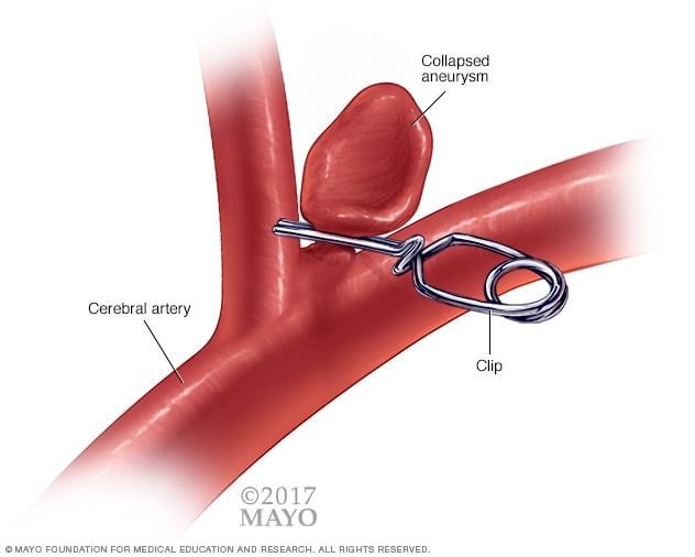
Surgical clipping is a procedure to close off an aneurysm. The neurosurgeon removes a section of your skull to access the aneurysm and locates the blood vessel that feeds the aneurysm. Then he or she places a tiny metal clip on the neck of the aneurysm to stop blood flow to it.
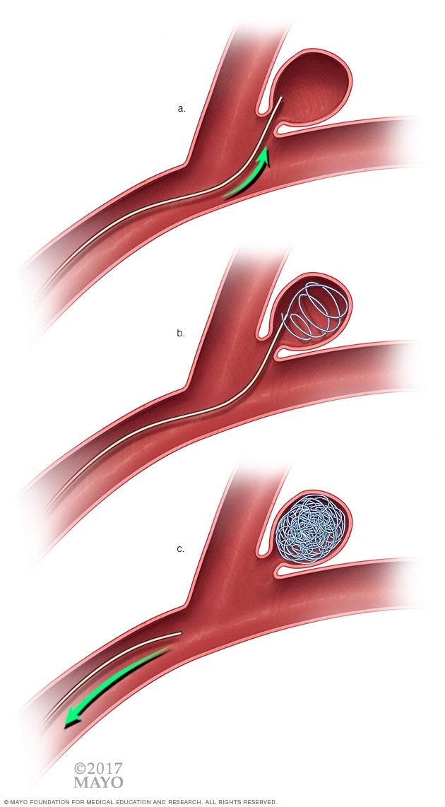
Endovascular coiling is a less invasive procedure than surgical clipping. The surgeon inserts a hollow plastic tube (catheter) into an artery, usually in your groin, and threads it through your body to the aneurysm. He or she then uses a guide wire to push a soft platinum wire through the catheter and into the aneurysm. The wire coils up inside the aneurysm, disrupts the blood flow and essentially seals off the aneurysm from the artery. This is what I had done.
Both procedures pose potential risks, particularly bleeding in the brain or loss of blood flow to the brain. The endovascular coil is less invasive and may be initially safer, but it may have a slightly higher risk of need for a repeat procedure in the future due to reopening of the aneurysm.
Flow diverters
Newer treatments available for brain aneurysm include flow diverters, tubular stent-like implants that work by diverting blood flow away from an aneurysm sac. The diversion stops blood movement within the aneurysm and so stimulates the body to heal the site, encouraging reconstruction of the parent artery. Flow diverters may be particularly useful in larger aneurysms that can’t be safely treated with other options.
Your neurosurgeon or interventional neuroradiologist, in collaboration with your neurologist, will make a recommendation based on the size, location and overall appearance of the brain aneurysm, your ability to undergo a procedure, and other factors.
Other treatments (ruptured aneurysms)
Other treatments for ruptured brain aneurysms are aimed at relieving symptoms and managing complications.
Pain relievers, such as acetaminophen (Tylenol, others), may be used to treat headache pain.
Calcium channel blockers prevent calcium from entering cells of the blood vessel walls. These medications may lessen the erratic narrowing of blood vessels (vasospasm) that may be a complication of a ruptured aneurysm. One of these medications, nimodipine (Nymalize, Nimotop), has been shown to reduce the risk of delayed brain injury caused by insufficient blood flow after subarachnoid hemorrhage from a ruptured aneurysm.
Interventions to prevent stroke from insufficient blood flow include intravenous injections of a drug called a vasopressor, which elevates blood pressure to overcome the resistance of narrowed blood vessels. An alternative intervention to prevent stroke is angioplasty. In this procedure, a surgeon uses a catheter to inflate a tiny balloon that expands a narrowed blood vessel in the brain. A drug known as a vasodilator also may be used to expand blood vessels in the affected area.
Anti-seizure medications may be used to treat seizures related to a ruptured aneurysm. These medications include levetiracetam (Keppra), phenytoin (Dilantin, Phenytek, others), valproic acid (Depakene) and others. Their use has been debated by several experts, and is generally subject to caregiver discretion, based on the medical needs of each patient.
Ventricular or lumbar draining catheters and shunt surgery can lessen pressure on the brain from excess cerebrospinal fluid (hydrocephalus) associated with a ruptured aneurysm. A catheter may be placed in the spaces filled with fluid inside of the brain (ventricles) or surrounding your brain and spinal cord to drain the excess fluid into an external bag. Sometimes it may then be necessary to introduce a shunt system — which consists of a flexible silicone rubber tube (shunt) and a valve — that creates a drainage channel starting in your brain and ending in your abdominal cavity.
Rehabilitative therapy. Damage to the brain from a subarachnoid hemorrhage may result in the need for physical, speech and occupational therapy to relearn skills.
Treating unruptured brain aneurysms
Aneurysm clip
Endovascular coiling
Surgical clipping or endovascular coiling or a flow diverter can be used to seal off an unruptured brain aneurysm and help prevent a future rupture. However, in some unruptured aneurysms, the known risks of the procedures may outweigh the potential benefit.
A neurologist, in collaboration with a neurosurgeon or interventional neuroradiologist, can help you determine whether the treatment is appropriate for you.
Factors to consider in making treatment recommendations include:
The size, location and overall appearance of the aneurysm
Your age and general health
Family history of ruptured aneurysm
Congenital conditions that increase the risk of a ruptured aneurysm
If you have high blood pressure, talk to your doctor about medication to manage the condition. If you have a brain aneurysm, proper control of blood pressure may lower the risk of rupture.
In addition, if you smoke cigarettes, talk with your provider about strategies to stop smoking since cigarette smoking is a risk factor for formation, growth and rupture of the aneurysm.
Mayo Clinic neurosurgeons are experts in each of these procedures and have pioneered many new techniques. They also regularly use advanced technology, such as 3D-printed models and computer simulations, to better understand the structure of the blood vessels and to plan surgery.
Mayo Clinic surgeons are trained in open vascular and endovascular neurosurgery, including minimally invasive techniques, such as the modified eyebrow incision, endoscopic skull base surgery and transnasal endoscopy. They are also experts in using computer-assisted technologies to navigate the brain during surgery and microvascular surgery.
Lifestyle changes to lower your risk
If you have an unruptured brain aneurysm, you may lower the risk of its rupture by making these lifestyle changes:
Don’t smoke or use recreational drugs. If you smoke or use recreational drugs, talk to your doctor about strategies or an appropriate treatment program to help you quit.
Eat a healthy diet and exercise. Changes in diet and exercise can help lower blood pressure. Talk to your doctor about changes appropriate for you.
Can you spot the 2 aneurysms? The big balloon was the largest and below it was the smallest one that is still intact. This is one of the pictures from my angio showing the aneurysm before the embolization. Zoom in close for details.This scan compares the brain pre-embolization (top half) and post-embolization (bottom half). Zoom in close for a clear view and details.
If you have experienced a severe headache or have any other symptoms related to a ruptured aneurysm your doctor will order tests to determine if blood has leaked into the space between the skull bone and brain.
Several tests are available to diagnose brain aneurysms and determine the best treatment. These include:
Computed tomography (CT). This fast and painless scan is often the first test a physician will order to determine if blood has leaked into the brain. CT uses x-rays to create two-dimensional images, or “slices,” of the brain and skull. Occasionally a contrast dye is injected into the bloodstream prior to scanning to assess the arteries, and look for a possible aneurysm. This process, called CT angiography (CTA), produces sharper, more detailed images of blood flow in the brain arteries. CTA can show the size, location, and shape of an unruptured or a ruptured aneurysm.
Magnetic resonance imaging (MRI). An MRI uses computer-generated radio waves and a magnetic field to create two- and three-dimensional detailed images of the brain and can determine if there has been bleeding into the brain. Magnetic resonance angiography (MRA) produces detailed images of the brain arteries and can show the size, location, and shape of an aneurysm.
Cerebral angiography. This imaging technique can find blockages in arteries in the brain or neck. It also can identify weak spots in an artery, like an aneurysm. The test is used to determine the cause of the bleeding in the brain and the exact location, size, and shape of an aneurysm. Your doctor will pass a catheter (long, flexible tube) typically from the groin arteries to inject a small amount of contrast dye into your neck and brain arteries. The contrast dye helps the X-ray create a detailed picture of the appearance of an aneurysm and a clear picture of any blockage in the arteries.
Cerebrospinal fluid (CSF) analysis. This test measures the chemicals in the fluid that cushions and protects the brain and spinal cord (cerebrospinal fluid). Most often a doctor will collect the CSF by performing a spinal tap (lumbar puncture), in which a thin needle is inserted into the lower back (lumbar spine) and a small amount of fluid is removed and tested. The results will help detect any bleeding around the brain. If bleeding is detected, additional tests would be needed to identify the exact cause of the bleeding.
I remember having a constant headache for 2 days or so prior to the rapture. The pain wasn’t overwhelming and I could tolerate it. I would rate it a 2 on the scale of 1-10 with 10 being the worst. New mothers, me being one of them, tend to ignore little things that their bodies are trying to communicate. We give excuses in order to make ourselves feel better. It is very true we are tired most of the time with little to no adequate rest most of the time. We prioritize the needs of our children and our loved ones. The question that still ponders my mind is – ‘was the aneurysm ruptured already prior to my hospital admission and the bleeding just got worse or did it rapture on that hectic night?’ The doctors that I have met with haven’t given me a conclusive answer yet. They tend to say…..in so many words——“we just don’t know”
So what are the actual symptoms of a brain aneurysm?
Please do not sit and start guessing what could be wrong with you or your loved one. Get emergency care if you suddenly get an intensely painful headache, lose consciousness, or have some of these other symptoms of an aneurysm that has ruptured: I cannot emphasize enough to you how critical it is to get that medical care urgently. Get help if you think something is wrong with your body. DO NOT WAIT.
Although brain aneurysms usually
don’t show symptoms, they can press on the brain and nerves as they get bigger.
See a doctor at once if you’re having the following symptoms of an unruptured
aneurysm:
Please remember that no matter what you are going through or how things turn out, you are not alone. God is with you and He has everything under control. He will lead you to where you need be at the right time. He will bring the right people to your case. God is watching us from a distance. You are a very special child of Him. Have faith and trust in nothing else but in Him. https://www.youtube.com/watch?v=hLHE9jrb_N4
Brain aneurysms/cerebral aneurysms form when the walls of the arteries in the brain become thin and weaken. Aneurysms typically form at branch points in arteries because these sections are the weakest. Occasionally, cerebral aneurysms may be present from birth, usually resulting from an abnormality in an artery wall. Reference
Risk
factors for developing an aneurysm
Sometimes
cerebral aneurysms are the result of inherited risk factors, including:
genetic
connective tissue disorders that weaken artery walls
polycystic
kidney disease (in which numerous cysts form in the kidneys)
arteriovenous malformations (snarled
tangles of arteries and veins in the brain that disrupt blood flow. Some
AVMs develop sporadically, or on their own.)
history
of aneurysm in a first-degree family member (child, sibling, or parent).
Other
risk factors develop over time and include:
untreated
high blood pressure
cigarette
smoking
drug
abuse, especially cocaine or amphetamines, which raise blood pressure to
dangerous levels. Intravenous drug abuse is a cause of infectious mycotic
aneurysms.
age
over 40.
Less
common risk factors include:
head
trauma
brain
tumor
infection
in the arterial wall (mycotic aneurysm).
Additionally,
high blood pressure, cigarette smoking, diabetes, and high cholesterol puts one
at risk of atherosclerosis (a blood vessel disease in which fats build up on
the inside of artery walls), which can increase the risk of developing a
fusiform aneurysm.
Risk
factors for an aneurysm to rupture
Not all
aneurysms will rupture. Aneurysm characteristics such as size, location,
and growth during follow-up evaluation may affect the risk that an aneurysm
will rupture. In addition, medical conditions may influence aneurysm rupture.
Risk
factors include:
Smoking.
Smoking is linked to both the development and rupture of cerebral aneurysms.
Smoking may even cause multiple aneurysms to form in the brain.
High
blood pressure. High blood pressure damages and weakens arteries, making
them more likely to form and to rupture.
Size.
The largest aneurysms are the ones most likely to rupture in a person who
previously did not show symptoms.
Location.
Aneurysms located on the posterior communicating arteries (a pair of arteries
in the back part of the brain) and possibly those on the anterior communicating
artery (a single artery in the front of the brain) have a higher risk of
rupturing than those at other locations in the brain.
Growth.
Aneurysms that grow, even if they are small, are at increased risk of rupture.
Family
history. A family history of aneurysm rupture suggests a higher
risk of rupture for aneurysms detected in family members.
The
greatest risk occurs in individuals with multiple aneurysms who have already
suffered a previous rupture or sentinel bleed.
For my case, I had been diagnosed
with high blood pressure in the first trimester of my second pregnancy and the
condition was being managed when I suffered the brain aneurysm. No family
history of brain aneurysms that I am aware of. No smoking, no alcohol abuse,
and absolutely no drug use except for the ones for BP and vitamins. My take
from this is that we are all pretty much at risk. I have heard stories of
completely healthy people, very athletic, and with no family history get them. The
more information we know about brain aneurysms, the more lives we can help
save.
Let’s be honest, how many people are familiar with brain
aneurysm? Even with a background in healthcare, I did not know what brain aneurysms
were all about except for the definition. Often, we find ourselves becoming
quite familiar with a condition once we experience it either directly or
indirectly.
Think of a weak spot in a balloon and how it feels stretched
out and thin. A brain
aneurysm is like that. It’s a weak spot in the wall of a blood vessel
inside the brain.
That area of the blood
vessel gets worn out from constant flow of blood and bulges out, almost like a
bubble. It can grow to the size of a small berry.
Although brain
aneurysms sound alarming, most don’t cause symptoms or health problems. You can
enjoy a long life without ever realizing that you have a brain aneurysm.
But in rare cases, aneurysms can grow big, leak, or explode.
Bleeding
in the brain, known as hemorrhagic stroke, is very serious and
requires urgent medical care.
A ruptured brain aneurysm can be life-threatening and lead
to:
cerebral vasospasm (reduced blood flow to the brain)
It’s in the raw hours of Saturday morning on July 29th, 2017, 1:00AM to be exact. Nothing much seems to be going on in this posh neighborhood of Kampala, except for the security guards struggling to stay awake. On the street of Naguru Vale, however, in one house, something is happening that will change the lives of this young aspiring family forever. “Somebody help, help me please, call the ambulance” A male voice is heard by the security guard on duty that night. Through the confusion, the security guard doesn’t know what do but listen keenly again for the voice. Meanwhile, the situation in this house is worsening. It’s a matter of life and death and time is of essence. “Post one, post one, can you read me? My wife is unresponsive, I need somebody to help me right now” My husband managed to get hold of the walkie talkie and placed a call that he hoped would end the nightmare.